Image:Testicle-histology-boar-2.jpg
From Wikipedia, the free encyclopedia
No higher resolution available.
Testicle-histology-boar-2.jpg (360 × 359 pixels, file size: 41 KB, MIME type: image/jpeg)
| | This is a file from the Wikimedia Commons. The description on its description page there is shown below.
|
| Description |
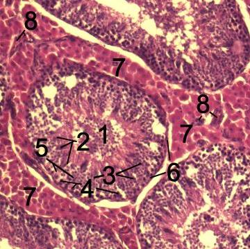
Histological section through testicular parenchyma of a boar. 1 Lumen of Tubulus seminiferus contortus, 2 spermatids, 3 spermatocytes, 4 spermatogonia, 5 Sertoli cell, 6 Myofibroblasts, 7 Leydig cells, 8 capillaries |
|---|---|
| Source |
Originally from en.wikipedia; description page is/was here. |
| Date |
2007-07-19 (original upload date) |
| Author |
Original uploader was Mikael Häggström at en.wikipedia |
| Permission (Reusing this image) |
CC-LAYOUT; CC-BY-SA-2.5,2.0,1.0; Released under the GNU Free Documentation License. |
[edit] License information
cellspacing="8" cellpadding="0" style="width:100%; clear:both; text-align:center; margin:0.5em auto; background-color:#f9f9f9; border:2px solid #e0e0e0;"

[edit] Original upload log
(All user names refer to en.wikipedia)
- 2007-07-19 15:13 Mikael Häggström 360×359×8 (41572 bytes) |Description=en:histological section through testicular parenchyma of a boar. 1 Lumen of Tubulus seminiferus contortus, 2 spermatids, 3 spermatocytes, 4 spermatogonia, 5 Sertoli-cell, 6 Myofibroblasts, 7 Leydig-cells, 8 capillaries<br> de: Histologisches
File history
Click on a date/time to view the file as it appeared at that time.
| Date/Time | Dimensions | User | Comment | |
|---|---|---|---|---|
| current | 09:20, 6 October 2007 | 360×359 (41 KB) | Kauczuk | ({{Information |Description=''no original description'' |Source=Originally from [http://en.wikipedia.org en.wikipedia]; description page is/was [http://en.wikipedia.org/w/index.php?title=Image%3ATesticle-histology-boar-2.jpg here]. |Date=2007-07-19 (origin) |
File links
The following pages on the English Wikipedia link to this file (pages on other projects are not listed):

