Image:Neuroblastoma mets.JPG
From Wikipedia, the free encyclopedia
No higher resolution available.
Neuroblastoma_mets.JPG (321 × 367 pixels, file size: 13 KB, MIME type: image/jpeg)
| | This is a file from the Wikimedia Commons. The description on its description page there is shown below.
|
| Description |
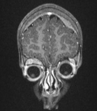
English: "This is a coronal post-gadolinium T1-weighted MPRAGE* MRI of a 2 year-old female with known neuroblastoma. There are orbital and skull vault metastases, with associated enhancing soft-tissue masses. The skull lesions are extradural masses which deform the underlying brain. The right orbital lesion forms a superior extraconal mass, depressing the right globe. Neuroblastoma not uncommonly metastasises to the skull. The metastases are generally osteolytic, enlarge the bone, strip the dura from it, and produce intracranial mass effect. Other skull metastases include osteogenic sarcoma (usually osteoblastic lesions); leukaemia; Wilm’s tumour; Ewing’s tumour; and hepatoblastoma." (dr Dawes) |
|---|---|
| Source | |
| Date |
25-2-2008 |
| Author |
Dr Laughlin Dawes |
| Permission (Reusing this image) |
author kindly mailed me permission to use this image outside cc-nc license; i mailed it forward to permissions-commons@wikimedia.org |
| This file is licensed under the Creative Commons Attribution 3.0 Unported License. In short: you are free to distribute and modify the file as long as you attribute its author(s) or licensor(s). Official license
Català | Česky | Deutsch | English | Ελληνικά | Español | Français | 한국어 | Italiano | עברית | Lietuvių | Magyar | Nederlands | Polski | Português | Русский | Türkçe | 中文(繁體) | +/- |
File history
Click on a date/time to view the file as it appeared at that time.
| Date/Time | Dimensions | User | Comment | |
|---|---|---|---|---|
| current | 19:19, 25 February 2008 | 321×367 (13 KB) | Filip em | ({{Information |Description=This is a coronal post-gadolinium T1-weighted MPRAGE* MRI of a 2 year-old female with known neuroblastoma. There are orbital and skull vault metastases, with associated enhancing soft-tissue masses. The skull lesions are extradu) |

