Image:Human brain left dissected midsagittal view description 2.JPG
From Wikipedia, the free encyclopedia
No higher resolution available.
Human_brain_left_dissected_midsagittal_view_description_2.JPG (701 × 488 pixels, file size: 50 KB, MIME type: image/jpeg)
| | This is a file from the Wikimedia Commons. The description on its description page there is shown below.
|
| Description |
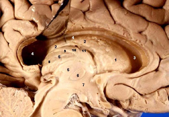
Human brain left dissected - midsagittal view Fornix & Septum Pellucidum Resected – Lateral Ventricle Exposed The head and body of the Caudate nucleus produce a large elevation in the lateral wall of the anterior horn and body of the lateral ventricle
|
|---|---|
| Source |
http://www.healcentral.org/healapp/showMetadata?metadataId=40566 |
| Date |
2005-11-30 |
| Author |
John A Beal, PhD Dep't. of Cellular Biology & Anatomy, Louisiana State University Health Sciences Center Shreveport |
| Permission (Reusing this image) |
CC-BY |
| Other versions | http://commons.wikimedia.org/wiki/Image:Human_brain_left_dissected_midsagittal_view.JPG |
[edit] Licensing
| This file is licensed under Creative Commons Attribution 2.5 License In short: you are free to distribute and modify the file as long as you attribute its author(s) or licensor(s). |
File history
Click on a date/time to view the file as it appeared at that time.
| Date/Time | Dimensions | User | Comment | |
|---|---|---|---|---|
| current | 22:14, 22 June 2006 | 701×488 (50 KB) | Patho | ({{Information| |Description='''Human brain left dissected - midsagittal view''' Fornix & Septum Pellucidum Resected – Lateral Ventricle Exposed The head and body of the Caudate nucleus produce a large elevation in the lateral wall of the anterior hor) |
File links
The following pages on the English Wikipedia link to this file (pages on other projects are not listed):

