Image:Gray219.png
From Wikipedia, the free encyclopedia
Size of this preview: 469 × 600 pixels
Full resolution (650 × 831 pixels, file size: 76 KB, MIME type: image/png)
| | This is a file from the Wikimedia Commons. The description on its description page there is shown below.
|
|
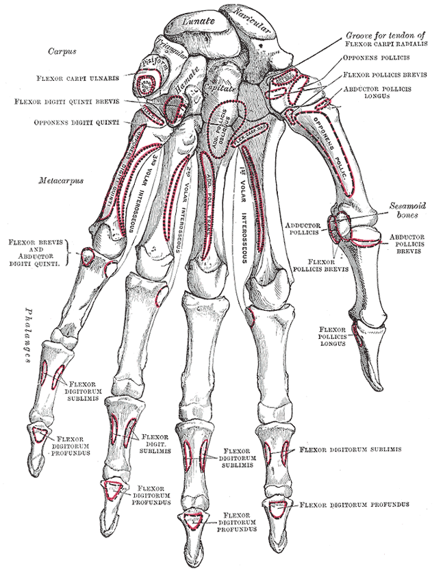
Caption: Bones of the left hand. Volar surface. This image is in the public domain because its copyright has expired. This applies worldwide. |
 |
File history
Click on a date/time to view the file as it appeared at that time.
| Date/Time | Dimensions | User | Comment | |
|---|---|---|---|---|
| current | 22:18, 17 March 2007 | 650×831 (76 KB) | Tene | (Optimised (0)) |
| 18:21, 13 April 2005 | 650×831 (78 KB) | Phyzome | ({{Gray's Anatomy plate| Bones of the left hand. Volar surface.}}) |
File links
The following pages on the English Wikipedia link to this file (pages on other projects are not listed):
- Carpus
- Gray's Anatomy
- Flexor digitorum profundus muscle
- Flexor digitorum superficialis muscle
- Image:The Obturator externus..gif
- Scaphoid bone
- Capitate bone
- Hamate bone
- Lunate bone
- Image:Gray1.png
- Wikipedia:Gray's Anatomy images with missing articles 5
- Image:Spleen1.png
- Image:Spleen2.png
- Image:Spleen3.png
- Image:Spleen4.png
- Image:Thorax-diaphragm.png
- Pisiform bone
- Triquetral bone
- Little finger
- Template:Gray's Anatomy plate
- Image:Grays anatomy leg 1.gif
- Image:Arm shoulder gray.gif
- Image:Prometaphase.jpg
- Image:Telophase.jpg
- Image:Left obicularis oculi.jpg
- Image:Levatores costarum.gif
- Image:Intertransversarii muscles.jpg
- Image:Vena saphena magna.gif
- Image:Small saphenous vein.gif
- Image:Anterior tibial a.gif
- Image:Axillary a.gif
- Image:Internal thoracic a.gif
- Image:Celiac a branches.gif
- Image:Superf veins of arm.gif
- Image:Svc ivc and azygos vs.gif
- Image:Lungs.gif
- Image:Mediastinum.gif
- Image:Vagus Glosso Acces course.gif
- Flexor carpi ulnaris muscle
- Flexor pollicis brevis muscle
- Flexor pollicis longus muscle
- Abductor pollicis longus muscle
- Opponens digiti minimi muscle
- Opponens pollicis muscle
- List of images in Gray's Anatomy: II. Osteology
- Flexor digiti minimi brevis (hand)
- Image:Telophase inverse.jpg
- Image:Pronator-teres.png
- Image:Gray1110-1.png
- Image:Internaliliac.png
- Image:Inferiorvesical.png
- Image:Midrectal.png
- Image:Mesoderm-mid.png
- Image:Maledevelopment.png
- Image:Gray1217-Castells Point-b.png
- Image:Radial sulcus.png
- Image:Scalenus.png
- Image:Gray411subclavius.png
- Image:Obliquus capitis inferior muscle.png
- Image:Obliquus capitis superior muscle.png
- Image:Rectus capitis posterior major muscle.PNG
- Image:Rectus capitis posterior minor muscle.png
- Image:Rectus capitis anterior muscle.PNG
- Image:Spinal column lateral.jpg
- Image:Infrahyoid muscles.PNG
- Image:Sternohyoid muscle.PNG
- Image:Thyrohyoid muscle.PNG
- Image:Suprahyoid muscles.PNG
- Image:Stylohyoid muscle.PNG
- Image:Mylohyoid muscle.PNG
- Image:Hyoglossal muscle.PNG
- User:Kelly/Image license templates

