Image:Cytokinesis-electron-micrograph.jpg
From Wikipedia, the free encyclopedia
No higher resolution available.
Cytokinesis-electron-micrograph.jpg (745 × 451 pixels, file size: 86 KB, MIME type: image/jpeg)
| | This is a file from the Wikimedia Commons. The description on its description page there is shown below.
|
Picture from English Wikipedia
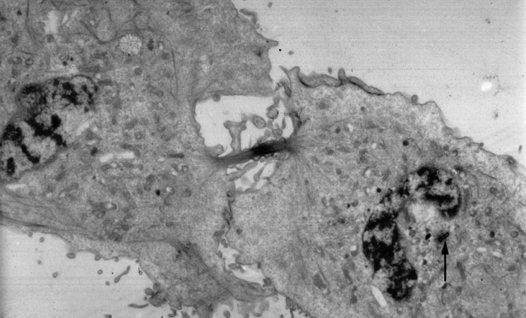
An electron micrograph image of a cell that has almost completed cell division and cytokinesis. Mitosis has already been completed. An arrow points to a centrosome still present near one of the nuclei.
From http://www.wadsworth.org/bms/SCBlinks/web_mit2/RES_MIT.htg/teleoph.jpg, the Wadsworth Center, which is part of the New York State Department of Health and devoted to public education. Since it's part of the US government, I'll assume public domain.
| This work is in the public domain in the United States because it is a work of the United States Federal Government under the terms of Title 17, Chapter 1, Section 105 of the US Code. See Copyright.
Note: This only applies to works of the Federal Government and not to the work of any individual U.S. state, territory, commonwealth, county, municipality, or any other subdivision. العربية | Български | Česky | Deutsch | English | Español | Français | Magyar | Italiano | 日本語 | 한국어 | Polski | Português | 中文(繁體) | 中文(简体) | +/- |
 |
Uploaded 07:21, 21 July 2005 .by user . Natalinasmpf . . 745x451 (87580 bytes) (An electron micrograph image of a cell that has almost completed cell division and cytokinesis. Mitosis has already been completed. An arrow points to a centrosome still present near one of the nuclei.
| This work is in the public domain in the United States because it is a work of the United States Federal Government under the terms of Title 17, Chapter 1, Section 105 of the US Code. See Copyright.
Note: This only applies to works of the Federal Government and not to the work of any individual U.S. state, territory, commonwealth, county, municipality, or any other subdivision. العربية | Български | Česky | Deutsch | English | Español | Français | Magyar | Italiano | 日本語 | 한국어 | Polski | Português | 中文(繁體) | 中文(简体) | +/- |
|
)
File history
Click on a date/time to view the file as it appeared at that time.
| Date/Time | Dimensions | User | Comment | |
|---|---|---|---|---|
| current | 17:43, 1 December 2005 | 745×451 (86 KB) | Rasbak | (Picture from English Wikipedia An electron micrograph image of a cell that has almost completed cell division and cytokinesis. Mitosis has already been completed. An arrow points to a centrosome still present near one of the nuclei. ) |

