Image:Bundleofhis.png
From Wikipedia, the free encyclopedia
No higher resolution available.
Bundleofhis.png (400 × 483 pixels, file size: 69 KB, MIME type: image/png)
| | This is a file from the Wikimedia Commons. The description on its description page there is shown below.
|
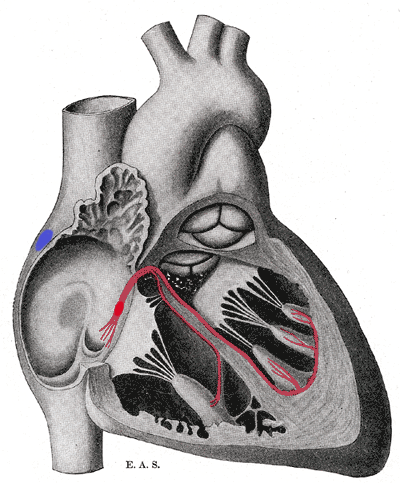
Schematic representation of the atrioventricular bundle of His. The bundle, represented in red, originates near the orifice of the coronary sinus, undergoes slight enlargement to form the AV node. The AV node tapers down into the bundle of HIS, which passes into the ventricular septum and divides into two bundle branches, the left and right bundles. Sometimes the 'left and right bundles of His' are called Purkyne or Purkinge fibres. The ultimate distribution cannot be completely shown in this diagram.
[edit] Licensing
|
|
This media file is in the public domain in the United States. This applies to U.S. works where the copyright has expired, often because its first publication occurred prior to January 1, 1923. See this page for further explanation.
|
|
|
Deutsch | English | Español | Français | Gaeilge | Galego | Italiano | Nederlands | 中文(简体) | +/- |
||
|
|
This image might not be in the public domain outside of the United States (this especially applies in Canada, China (not Hong Kong, Macao, or Taiwan Area), Germany, Mexico, and Switzerland). The creator and year of publication are essential information and must be provided. See Wikipedia:Public domain and Wikipedia:Copyrights for more details. |
File history
Click on a date/time to view the file as it appeared at that time.
| Date/Time | Dimensions | User | Comment | |
|---|---|---|---|---|
| current | 17:40, 20 September 2006 | 400×483 (69 KB) | Kauczuk | (Bundle of His, from Gray's Anatomy 1918) |
File links
The following pages on the English Wikipedia link to this file (pages on other projects are not listed):

